Brain activity can be recorded in vivo using several non-invasive techniques, such as functional magnetic resonance imaging (fMRI), electroencephalography (EEG), and magnetoencephalography (MEG). These techniques are intrinsically complementary because if they present a high spatial resolution, they also lack in time resolution, i.e. fMRI, and vice versa, i.e. EEG and MEG. fMRI is able to identify the main cognitive and sensorimotor circuits, i.e., resting state network, of the brain but it’s not able to detect the spontaneous brain waves characteristic of different awareness states, which can be instead provided by EEG/MEG recordings. In this context, it’s easy to understand how a multimodal approach is needed in order to deepen our knowledge of the neural bases of brain activity.
A multimodal approach is useful also to improve brain modelling findings in pathological conditions, especially in heterogeneous clinical conditions, such as mild cognitive impairment (MCI). Indeed, brain modelling is a promising approach that not only integrates knowledge across various scales but also uses high temporal resolution signals to constrain and drive realistic brain activity simulations in order to obtain personalized profiling for healthy and pathological subjects and disease stratification.
A PhD student will have the possibility to cover various aspects of the investigation of brain functioning with the aim of identifying strategies for diagnosis and personalized therapy. Students have the possibility to spend part of their research activity abroad since the projects are developed within a collaborative network, which comprises MNESYS, CN1, EBRAINS 2.0, and Virtual Brain Twin (VBT) projects.

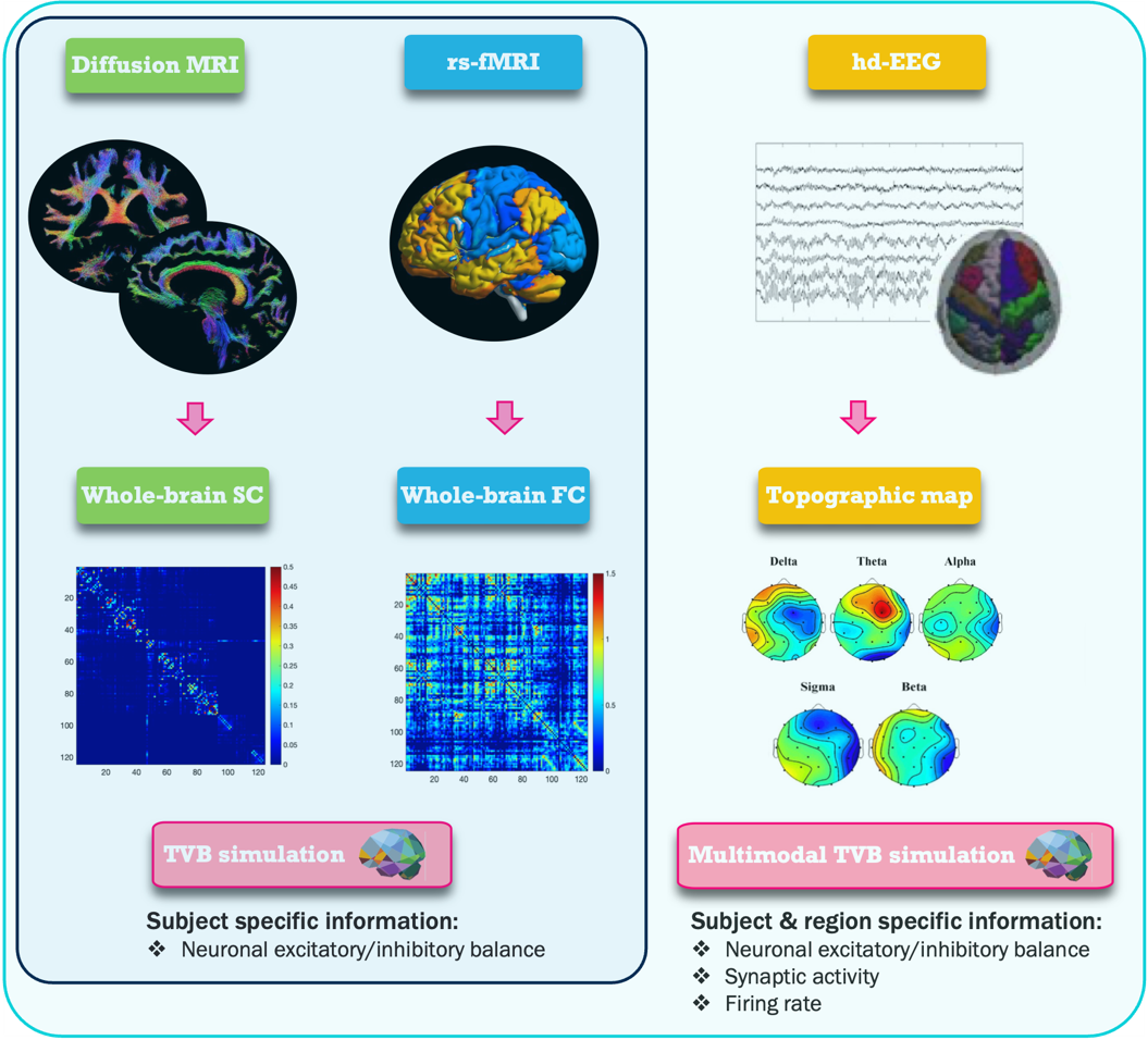
Multimodal virtual brain modelling.
Brain connectivity is used as input to The Virtual Brain (TVB), while functional connectivity is used as target for model tuning.
TVB identifies a subject-specific excitatory/inhibitory balance based on the optimal value of the tuned parameters.
Multimodal TVB uses EEG recordings to constrain input data and drive simulation in order to obtain a detailed subject and region specific description.
